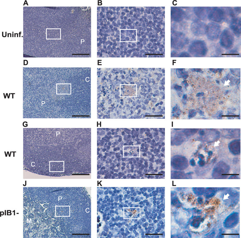
Figure 3. Yptb Localizes in the Cortex and Paracortex of the MLN.
BALB/c mice were intragastrically inoculated with 2 × 10⁹ WT or IP2666 pIB1−. At 4 d post-inoculation, MLN were harvested and stained for Yptb followed by hematoxylin staining. Picture sections of uninfected (A–C), WT infected (D–I), and pIB1− infected (J–L) MLN were taken at 150×, 900×, and 4,500× magnification. White boxes indicate magnified areas in the next slide. Arrows point to Yptb microcolonies. Scale bars correspond to 133 μm for 15× magnification, 22 μm for 90× magnification, and 4.4 μm for 450× magnification. Pictures shown are representative of multiple fields and samples from MLN infected with WT or pIB1−.
C, cortex; gc, germinal center; M, medulla; P, paracortex.

