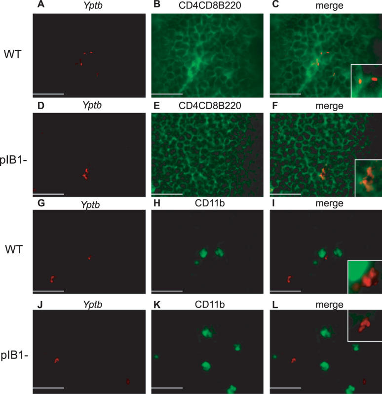
Figure 4. pTTSS Mutants Are Adjacent to B and T Lymphocytes Whereas WT Is Adjacent to B and T Lymphocytes and CD11b+ Cells.
BALB/c mice were intragastrically inoculated with 2 × 10⁹ WT IP2666 or IP2666 pIB1−. At 4 d post-inoculation, MLN were harvested, sectioned, and examined by fluorescence microscopy. Staining with antibodies to Yptb (red) (A), (D), (G), and (J), to CD4-CD8-B220 (green) (B) and (E), or to CD11b (green) (H) and (K) was performed, and images merged (C), (F), (I), and (L). Pictures show the cortex–paracortex and are representative of multiple fields and samples for WT-infected mice and pIB1 mice stained with CD4-CD8-B220. Fewer fields had pIB1 bacteria and CD11b+ cells. Scale bars correspond to 22 μm for 900× magnification.

