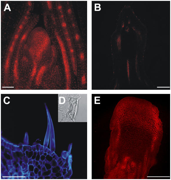
Figure 8.
ZmPIN1 localization in bif2 vegetative and reproductive tissues. The bif2 mutant presents wild-type vegetative development, but abnormal reproductive structures. During the vegetative phase, bif2 plants develop a normal phyllotaxis and the putative auxin efflux carrier localization appears the same as in wild-type maize. After the switch to the reproductive phase, the tassel and ear show several developmental defects and PAT seems to be completely impaired. A, Longitudinal section of a bif2 SAM, where ZmPIN1 presents a wild-type localization. The 7-d after germination bif2 SAM section was labeled with the anti-AtPIN1 antibody coupled to an Alexa568 fluorochrome (red). B, In the fully developed tassels of severe bif2 mutants, the ZmPIN1 is completely absent. This image shows a longitudinal tassel section, which presents no polar signal; tissue autofluorescence appears red. C, Epifluorescence image of a longitudinal section detail of a mutant tassel at flowering. Structures appear blue as autofluorescence was observed with a 4′,6-diamino-phenylindole filter. In the tassel of severe bif2 mutants, the antibody does not recognize any membrane protein. The male inflorescence presents no AMs and a completely disrupted vasculature. The developmental defects include the production of leaf-like epidermis, which carries stomata (D) and trichomes. E, Epifluorescence image of a bif2 adult ear. Red color codes for the Alexa568 fluorochrome. In female reproductive structures, AMs are absent and the vascular bundles are disrupted, although the defects seem less severe than in the tassel. The anti-PIN1 antibody labels polarly localized proteins in the vasculature. Bars: A = 100 μm; B and E = 75 μm; C = 30 μm.

