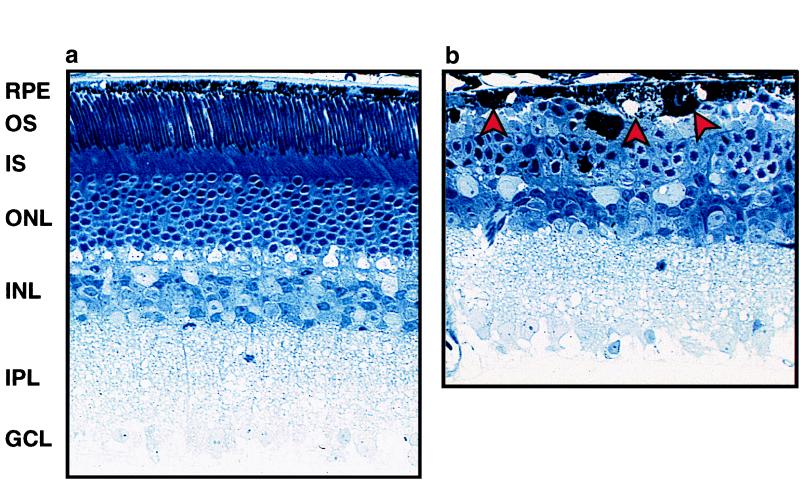
Figure 5.
Retinal histopathology of Oat−/− mice on an arginine-restricted diet (a) or a standard diet (b) at age 12 months. Note the reduced number of ONL cells and loss of IS and OS in the right panel. The arrows indicate abnormal RPE cells. GCL, ganglion cell layer; IPL, inner plexiform layer; INL, inner nuclear layer; ONL, outer nuclear layer; IS and OS, inner and outer segments, respectively, of photoreceptors.

