
Fig. 3.
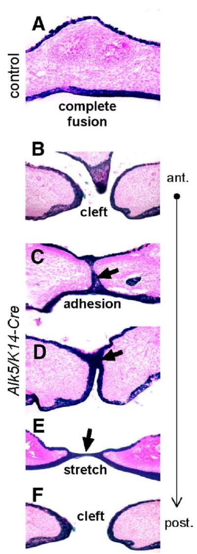
Fate mapping of midline epithelial cells in Alk5/K14-Cre mutants. Transverse palatal sections of E17 embryos expressing K14-Cre in the Rosa26 reporter background were stained for β-galactosidase activity (blue), which permanently marks epithelial cells and their descendants (eosin counterstain, magnification 20×). (A) Control palates have cells derived from the epithelium exclusively on the oral and nasal side of the palate, whereas the fully confluent mesenchyme (pink) along the entire anterior–posterior axis is devoid of positively staining cells. (B–F) Alk5/K14-Cre mutants show either no adhesion (cleft) or adhesion with a lack of fusion detected as persistent midline epithelial seam in the anterior palate (blue staining and arrows). Moreover, the nasal septum has failed to fuse with palatal shelves (B). The posterior part of the palate shows a stretched epithelial bridge (E), followed by palatal cleft in the most posterior region of the soft palate (F).