
Fig. 7.
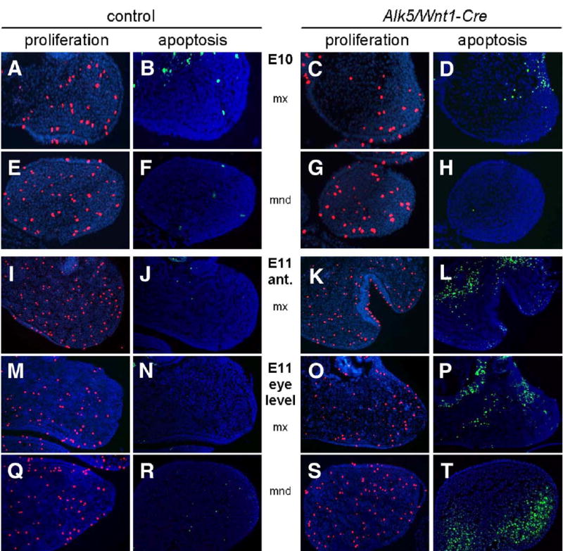
Increased apoptosis in pharyngeal arches of Alk5/Wnt1-Cre mutants. Frontal sections show a pronounced increase in number of TUNEL-positive apoptotic cells (green signal) in the maxillary (mx) and mandibular (mnd) processes of Alk5/Wnt1-Cre mutants on E11, but not on E10, when compared to controls. Cell proliferation (anti-phosphohistone H3 immunostaining, red signal) is comparable between mutants and controls (blue counterstaining with DAPI; magnification 20×).