
Fig. 9.
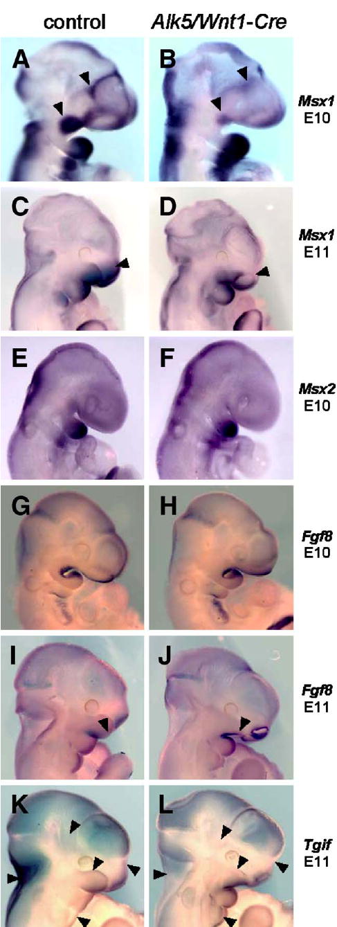
Attenuated expression of Msx1 and changes in expression patterns of Fgf8 and Tgif in Alk5/Wnt1-Cre mutants. Endogenous gene expression was visualized by in situ hybridization with biotinylated riboprobes (blue signal, magnification 5× for E10 and 4× for E11 samples). Whereas Msx2 did not show any change, Msx1 shows a remarkable decrease in expression in the maxillary process and frontonasal mass in mutants at E10 (B, arrowheads) and in the upper nasal pit region at E11 (D, arrowhead). Fgf8 expression in Alk5/Wnt1-Cre mutants spreads more anteriorly along the lower edge of the maxillary process at E11 (J, arrowhead), when compared with the control (I). Tgif expression shows a slight increase in the mandibular process and the 2nd pharyngeal arch in mutants at E11, whereas its expression in mutants of the same age is attenuated or absent in the upper portion of the maxillary process of the 1st pharyngeal arch, frontonasal process, temporoparietal region, and the nuchal area (K–L, arrowheads) The nasal pits and most of the calvaria show a comparable staining pattern and intensity in both mutants and controls.