
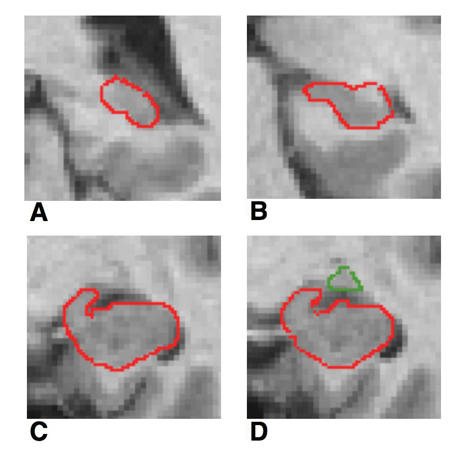
Fig. 1: Coronal MRI slices that run in occipito-rostral direction: (A) most posterior coronal slice where the hippocampus was clearly detectable, (B) hippocampal body; (C) the shape of the hippocampus may be compared with a rabbit with the head vertically directed, (D) amygdala-hippocampal transition area (HATA).