Figure 1.
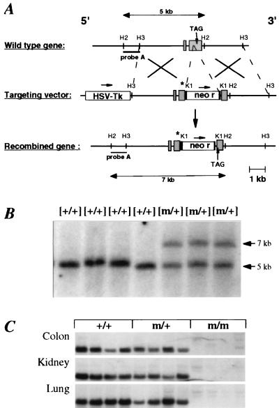
Gene targeting strategy for the disruption of the mouse βENaC gene locus. (A) Structure of the wild-type βENaC gene, the targeting vector, and the predicted targeted locus. The “R566Stop” mutation is indicated (asterisk). Expected fragment sizes of the wild-type and mutated βENaC allele after digestion with HincII and hybridization with probe A are indicated. H2 = HincII; H3 = HindIII; K1 = KpnI. The precise exon–intron structure has not been determined. Identified exons are indicated (shaded box). (B) Southern blot analysis of offspring from the breeding of chimeras. Tail DNA was digested with HincII and hybridized with a 5′ flanking probe (probe A). The βENaC genotype is indicated above each lane. Expected fragment sizes of wild-type (5-kb) and mutant (7-kb) allele are indicated. (C) Northern blot analysis of βENaC RNA transcripts from βENaC +/+, m/+, and m/m mice kept on low-salt diets (1 g/kg Na+). Total RNA (15 μg) from colon, kidney, and lung of each four independent animals was hybridized with βENaC-specific 32P-labeled probes.

