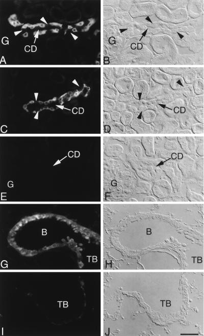
Figure 2.
Immunolocalization of βENaC protein in kidney (A–F) and lung (G–J) of βENaC +/+ and βENaC m/m mice by using C-terminal antibody (A and B) and ectodomain antibody (C–J). (Left) Immunofluorescence. (Right) Corresponding Normarski images. In cortical collecting ducts (CD) from wild-type mice (A–D), immunofluorescence was present in the cytoplasm of principal cells, whereas intercalated cells (arrowheads) were unreactive. Cells of proximal convoluted tubules and glomeruli (G) were unstained. In contrast, no staining was detected in kidney of βENaC m/m mice (E and F). In lung of wild-type mice (G and H), signal was present in bronchioles (B) and terminal bronchioles (TB). No specific immunostaining was present in lung from the βENaC m/m mice (I and J). The faint, nonspecific fluorescence in I is nonepithelial and associated with basement membrane. (Scale bar = 25 μm.)

