Figure 2.

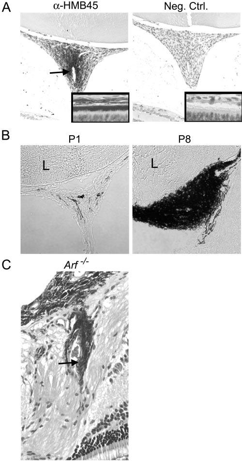
RPE-like cells accumulate in retrolental fibrovascular mass in Arf−/− mouse. (A) Representative photomicrographs of retrolental tissue from P10 albino Arf−/− mouse stained with α-HMB45 antibody (left) and unrelated primary antibody (right). HMB45-positive cells in retrolental tissue localize around large blood vessel (arrow). Insets: antibody staining of RPE as control. (B) Phase contrast photomicrographs of cryostat sections through retrolental tissue showing accumulation of pigmented cells between P1 and P8 in Arf−/− mice. (C) Photomicrograph of hematoxylin and eosin–stained section shows pigmented cells adjacent to vascular structures (arrow) within retina near the optic nerve/hyaloid artery stalk in 3-month-old Arf−/− mouse. Original magnification: (A, B) ×200 and ×400 (insets in A; C).
