Figure 1.

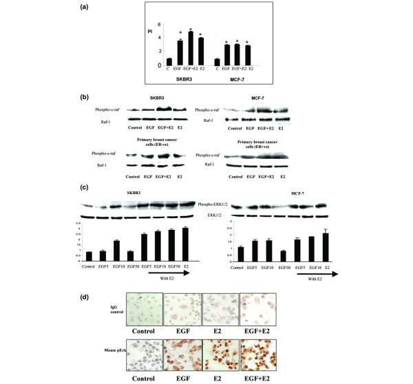
Effect of 17β-oestradiol and EGF on cell proliferation and induction of MAPK protein expression in breast cancer cells. (a) Cell proliferation findings. SKBR3 and MCF-7 human breast cancer cells, and ER-negative and ER-positive primary breast cultures derived from patient tumours were treated with EGF (10 ng/ml) and 17β-oestradiol (10-8 mol/l) alone and in combination for 24 hours. Cell proliferation assays were carried out using MTT thiozolyl blue. Proliferative index of the control group is standardised to 1. The results shown are expressed as mean ± standard error (n = 9). Statistical analysis was performed using Mann Whitney U test (*P < 0.02 versus control). (b) Western blot analysis of phospho-Raf and total Raf protein expression. SKBR3 and MCF-7 human breast cancer cells, and ER-negative and ER-positive primary breast cultures derived from patient tumours were treated with EGF (10 ng/ml) and 17β-oestradiol (10-8 mol/l) alone and in combination for 10 minutes. (c) Phospo-ERK1/2 and total ERK1/2 protein expression. SKBR3 and MCF-7 human breast cancer cells were treated with 5, 10 and 50 ng/ml EGF and 17β-oestradiol (10-8 mol/l) alone and in combination for 10 minutes. Results are representative of those obtained in three separate experiments. (d) Immunocytochemical localisation of phospho-ERK in ER-negative SKBR3 cells treated with EGF (10 ng/ml) and 17β-oestradiol (10-8 mol/l) alone and in combination for 30 minutes. The slides were counter stained with Mayer's haematoxylin solution and viewed under a light microscope at 200× magnification. Positive cells stained cells stained brown against a blue background. Results are representative of those obtained in three separate experiments. E2, 17β-oestradiol; EGF, epidermal growth factor; ER, oestrogen receptor; ERK, extracellular signal regulated kinase; MAPK, mitogen-activated protein kinase; PI, proliferative index.
