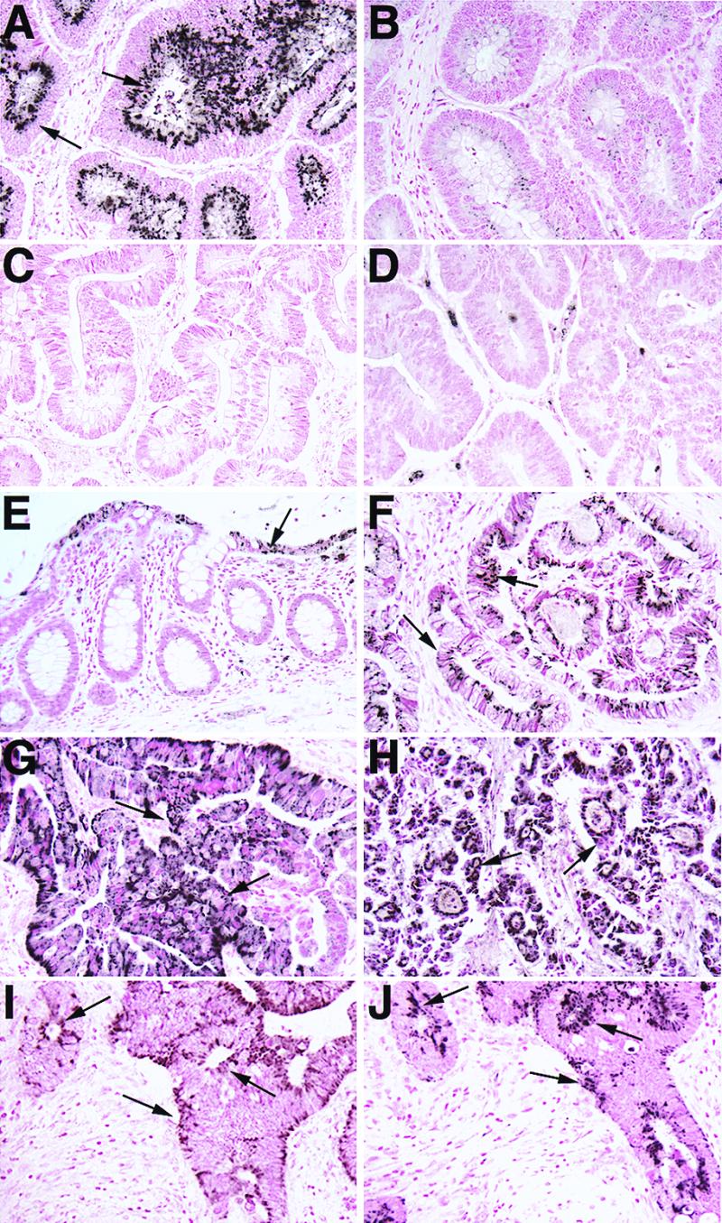
Figure 2.

IHC of M68 and FAS in normal and tumor tissues of GI tract. Note strong positive staining in malignant epithelial cells (arrows) in tumor samples (A, colon; F, esophagus; G, stomach; and H, rectum). In normal adjacent colon (E), weak M68 expression was detected in epithelial cells (arrows) lining the lumen and was generally absent in the glandular epithelium. A significant decrease in staining was observed when M68 antibody was preincubated with the immunizing peptide (B), and no tumor epithelial cell staining was observed in tumor tissues with preimmune serum (C) or a nonimmune rabbit serum (D). IHC of Fas (CD95) (I) and M68 (J) in a colon adenocarcinoma. Note the similar staining pattern and coexpression of CD95 and M68 in the tumor epithelial cells (arrows).
