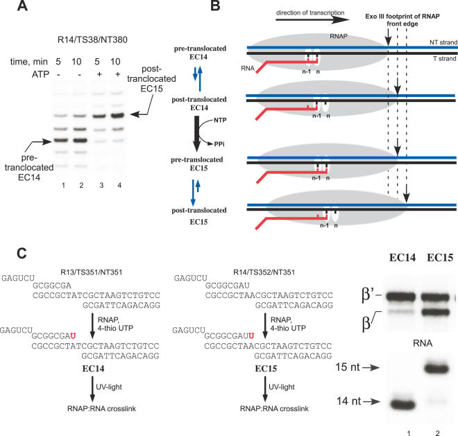
Figure 2.
Probing of the EC translocation conformations. (A) Exo III footprints of EC14 and EC15. Tth EC14 (lanes 1 and 2) was assembled as described in Materials and Methods. EC15 (lanes 3 and 4) was obtained by incubation of EC14 with substrate ATP for 2 min at 60°C. Exo III (0.02 U/μl) was added for 5 (lanes 1 and 3) or 10 (lanes 2 and 4) min at 37°C. (B) Schematics of RNAP front edge oscillations in EC14 and EC15. (C) Photo cross-linking patterns of Tth EC14 and EC15, EC14 and EC15 containing 5′ 32P-labeled RNA primers were prepared as illustrated (left). The photo cross-linking analog 4-thio UTP (50 μM) was incorporated into the transcript for 2 min at 60°C followed UV light irradiation for 5 min at RT. The cross-linked species were separated using gel electrophoresis (right).

