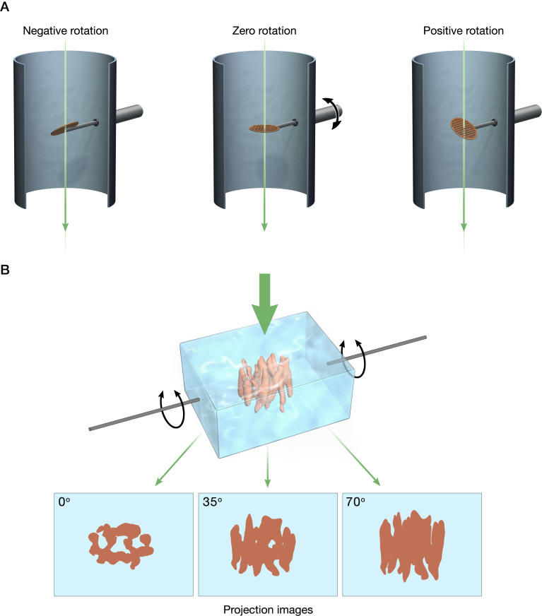
Figure 1. Principle of 3-D Imaging Using Electron Tomography.
(A) Schematic illustrating the principle of data collection for electron tomography by tilting specimens relative to the electron beam.
(B) Rendering of three “perfect” projection views (at 0°, 35°, and 70°) generated by rotating a vitrified film of ice-embedded molecules relative to the electron beam. The structure shown is that of the oxalate transporter determined at 6.5 Å resolution using electron crystallography [18]. Structure determination by electron tomography involves starting with a set of projection images which are then effectively “smeared” out along their viewing directions to form back-projection profiles. These profiles are combined appropriately to recover the density distribution of the imaged object.

