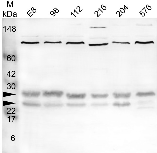
Figure 3.
Western blot probed with an affinity purified antibody, anti-MrgR, showing the detection of MrgR in whole cell lysates from 6 isolates of B. pseudomallei grown at 37°C. The isolate number is indicated above each lane. Lane M: molecular weight markers indicated in kilodaltons (kDa). See Table 1 for isolate details. Arrows indicate the expected position of the 24 kDa MrgR protein and slightly larger phosphorylated forms of MrgR. See text for details.

