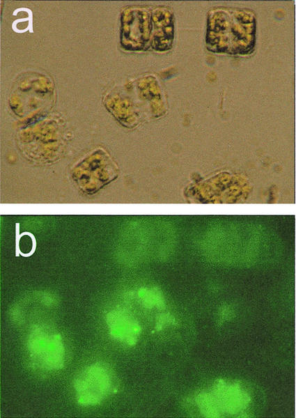
Figure 4.
Epifluorescense microscopy of intact and disrupted T. rotula treated with the fluorogenic substrate 1,2-bis-(BODIPY-3-undecanoyl)-sn-glycero-3-phosphocholine. A, Light microscopic control of the cells (left, intact cell; right, cell damaged during preparation). B, Same part, excited with UV light: λex = 450 to 490 nm and λem = 515 to 565 nm. The green fluorescence is due to the presence of lysolipids after action of T. rotula PLA2.

