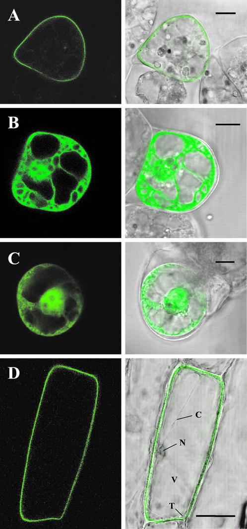
Figure 7.
Subcellular localization of GFP fusion proteins. Wild-type LeCPK1 (A) and the G2A site-directed mutant of LeCPK1 (B) were transiently expressed as C-terminal GFP-fusion proteins in suspension-cultured L. peruvianum cells. Cells in C were transformed to express GFP alone. D, Onion epidermal cells expressing the wild-type LeCPK1-GFP fusion. Letters indicate the nucleus (N), the vacuole (V), the tonoplast (T), and a cytoplasmic strand (C). The localization of GFP-fusion proteins was analyzed by confocal laser scanning microscopy (left). Merged pictures of the green fluorescence channel with the corresponding light micrographs are shown on the right. The length of the bars corresponds to 10 (A–C) and 50 (D) μm, respectively.

