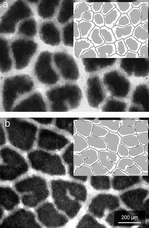
Figure 1.
The abaxial surface of an intact leaf of Q. coccifera illuminated from the adaxial surface and viewed with a light microscope under low magnification. a, Sun-exposed leaf. b, Shade-exposed leaf. The inserts show the particular leaf area classes segmented by the image analysis procedure (see “Materials and Methods”). White region, At; gray region, Ap; solid line, trace of BSEs. The values of the particular parameters for these samples were: a, leaf thickness of 0.37 mm; Ap 54.1%; λt 0.084 mm3 mm−2; and CFAt 345 mm mm−2; b, leaf thickness of 0.25 mm; Ap 73.6%; λt 0.031 mm3 mm−2; CFAt 210 mm mm−2.

