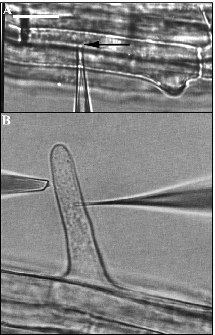
Figure 7.
Microphotgraphic examples of pressure probe (A) and ion-flux/voltage-clamp measurements (B). The micropipette tip is indicated by an arrow in A. It is located in the vacuole. In B, the double-barreled microelectrode is impaled into the cytoplasm (Lew, 2000). The ion fluxes from the root hair were measured parallel to the root surface. Bar = 20 μm.

