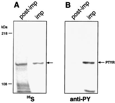
Figure 1.
Radioactive profile of tyrosine-phosphorylated proteins after infection of AGS cells with 35S-labeled Hp. AGS cells were infected with the 35S-labeled Hp strain 87A300. Tyrosine-phosphorylated proteins were immunoprecipitated from RIPA-buffer-soluble fractions by using monoclonal anti-phosphotyrosine antibody (anti-PY; PY20, Transduction Laboratories, Lexington, KY). Immunoprecipitates (imp) and proteins of the RIPA-soluble cell fraction after the immunoprecipitation with anti-PY (post-imp) were separated by SDS/6% PAGE and transferred onto nitrocellulose membranes in duplicate. One membrane was exposed to Kodak x-ray film (A); the second nitrocellulose membrane was probed with anti-phosphotyrosine antibody (anti-PY). Blots were developed with peroxidase-coupled secondary antibodies [GIBCO/BRL]. The arrow in A indicates the 35S-labeled protein immunoprecipitated with anti-PY and comigrating with PTYR (shown in B).

