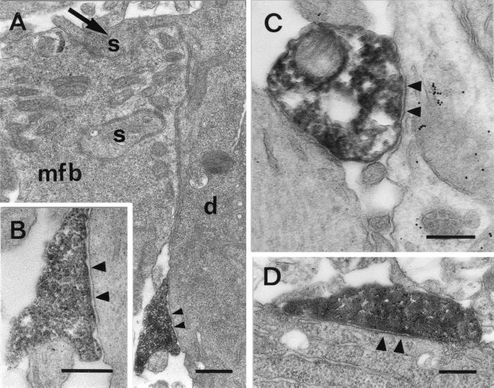
Figure 2.
Synapses formed by MFA interneurons in CA3. (A) Synaptic contact (◂) on a proximal apical dendritic shaft (d) made by a biocytin-filled bouton of an identified MFA interneuron. The dendrite was identified as belonging to a pyramidal cell by the large spines (s) establishing synaptic contacts with a mossy fiber bouton (mfb); one of the spines (arrow) is in direct connection with the dendritic shaft. (B) Higher magnification of the symmetric synaptic contact (◂) shown in A. (C) Biocytin-filled bouton of an MFA cell establishing symmetric synaptic contact (◂) with a spinefree, beaded dendrite. This dendrite most likely belongs to a GABAergic inhibitory neuron as indicated by the postembedding GABA immunogold labeling. (D) Symmetric synaptic contact (◂) formed by a biocytin-filled bouton on a cell body in the pyramidal layer of CA3. (Bars = (A) 0.5 μm; (B) 0.3 μm; and (C and D) 0.25 μm.)

