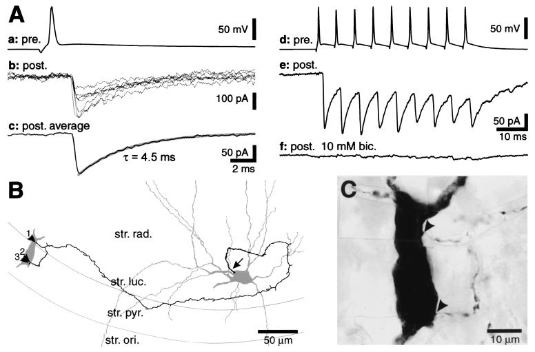
Figure 3.
Postsynaptic effect of an MFA interneuron on a morphologically identified CA3 pyramidal cell. (A) APs in the presynaptic interneuron (Aa) were followed by IPSCs of short latency in the postsynaptic pyramidal cell [(Ab) seven consecutive IPSCs superimposed; (Ac) average of 12 IPSCs; membrane potential −70 mV]. The decay phase of the average IPSC could be fitted with a single exponential with a time constant (τ) of 4.5 ms. During trains of APs (Ad), IPSCs showed a constant peak amplitude in the averaged response (n = 26, Ae). Bath perfusion of bicuculline (bic., 10 μM) blocked the synaptic response (Af, average of 25 traces, between 1.5 and 3 min of drug wash-in). (B) Camera lucida reconstruction of the presynaptic MFA interneuron (on the right) and the postsynaptic pyramidal cell (soma and proximal dendrites depicted only) with the route of the axon to the boutons forming the synaptic contacts. For clarity, only this part of the axonal plexus is illustrated. The arrow points to the axon initial segment; arrowheads indicate the boutons (1–3) forming synaptic contacts with the pyramidal cell. Bouton 3 was not observed in the light microscope but was found in the electron microscope when analyzing the serial thin sections. (C) Photomontage of the section containing the soma of the biocytin-filled postsynaptic pyramidal cell. The synaptic contacts identified in the light microscope are indicated by arrowheads. str. ori., stratum oriens; str. pyr., stratum pyramidale; str. luc., stratum lucidum; and str. rad., stratum radiatum.

