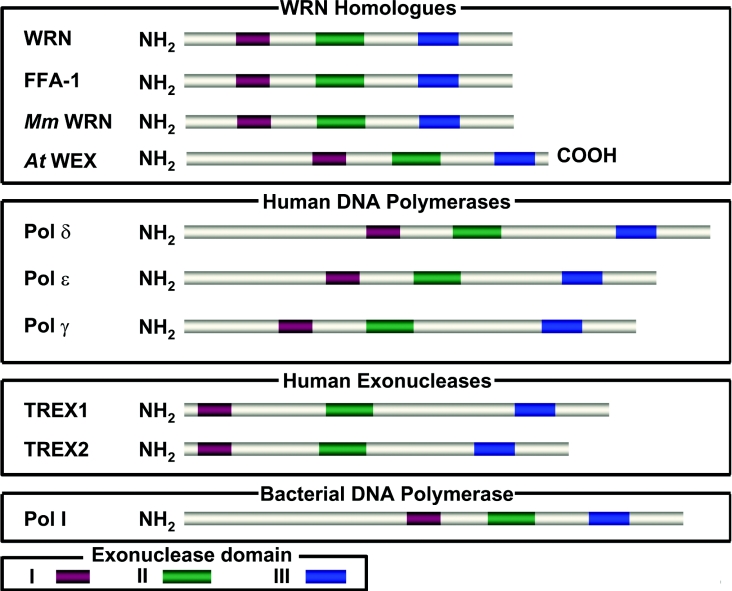
Figure 2. Conserved nuclease domain of WRN and 3′ to 5′ exonucleases.
The top panel depicts the three conserved exonuclease motifs of human WRN and its homologues in lower eukaryotes. The two middle panels depict human DNA polymerases and exonucleases which share identity with WRN exonuclease. The bottom panel depicts a prokaryotic DNA polymerase with the conserved exonuclease domains. The conserved exonuclease domains are colour-coded as indicated at the bottom. Mm, Mus musculus.

