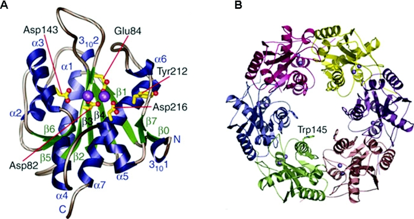
Figure 4. WRN exonuclease structure.
(A) X-ray crystal structure of the WRN-exo fold is shown with the conserved active site residues that chelate the two Mn2+ ions (purple) [35]. (B) The WRN-exo hexameric ring model with uniquely coloured WRN-exo subunits was built by superimposition with the A. thaliana homologue [35]. Figures are courtesy of Dr John Tainer (The Scripps Research Institute, La Jolla, CA, U.S.A.).

