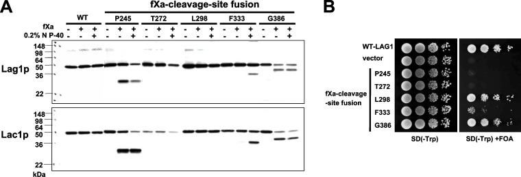
Figure 3. Topology determination by insertion of protease cleavage sites.
(A) fXa protease cleavage of Lag1–fXa and Lac1–fXa fusion proteins. RH382 cells were transformed with plasmids 1810–1819 to express the Lag1–fXa and Lac1–fXa fusion proteins. The numbers and amino acids represent the amino acid residues of Lag1p and Lac1p before which the fXa protease cleavage site was inserted. The microsomes were mock-digested or digested with fXa protease in the absence or presence of NP40 on ice for 2 h, and resolved on 13.5% polyacrylamide gels. The Lag1 and Lac1 proteins were detected by immunoblotting using anti-FLAG antibodies. (B) Functional complementation of lag1Δ lac1Δ cells by Lag1–fXa cleavage site fusion proteins. lag1Δ lac1Δ double deletion mutant cells transformed with pRS416-LAG1 (LAG1 on uracil-based plasmid) (RH6602) were transformed with pRS414-LAG1-fXa4-8 (LAG1-fXa fusion constructs on tryptophan-based plasmid) (plasmids 1820–1824). The transformants were tested for complementation as in Figure 2(B).

