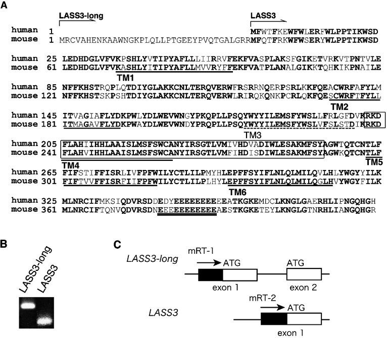
Figure 1. Amino acid sequence of mouse LASS3.
(A) Comparison of the mouse and human LASS3 amino acid sequences (GenBank™ accession numbers, DQ646881/DQ358087 and BC034970, respectively) is shown. LASS3-long contains 36 additional amino acid residues at the N-terminus. Conserved amino acids are shown in bold. The Lag1 motif is enclosed by a box. The clusters of Glu residues, characteristic of LASS3 proteins, are indicated by double underlining. The TopPredII program [18] predicted that LASS3 comprises up to six TM spanning domains, shown here underlined. TM1, TM2, TM4 and TM5 each exhibited high probability scores (TM1, 1.1948; TM2; 1.4104, TM4, 1.3427; TM5, 1.8896), whereas the scores for TM3 and TM6 were relatively low (1.0630 and 1.0260 respectively). Taking the topology model of LASS6 [9] into consideration, we propose that only five domains, TM1, TM2, TM4, TM5 and TM6 (continuous underlining) span the membrane and that TM3 (broken underlining) does not. (B) The mRNA expression of LASS3 and LASS3-long was determined by RT-PCR using mouse testis cDNA as detailed in the Materials and methods section. (C) The first and second exons of LASS3-long, the first exon of LASS3, and the primers used in (B), are shown.

