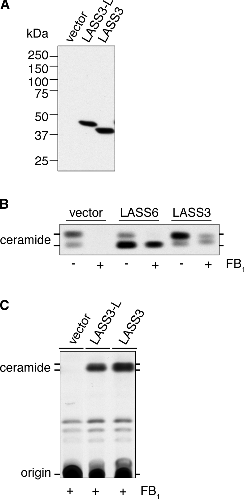
Figure 2. (Dihydro)ceramide synthase activity of LASS3.
HEK-293T cells were transfected with pcDNA3-HA (vector), pcDNA3-HA-LASS3 (LASS3), pcDNA3-HA-LASS3-L [LASS3-long (LASS3-L)] or pcDNA3-HA-LASS6 (LASS6) as indicated. (A) Total cell lysates (5 μg of protein) were separated on an SDS/10%-(w/v)-polyacrylamide gel then transferred to a membrane. Immunoblotting was then performed using an anti-HA antibody. (B) Transfected cells were incubated with 1 μCi of [3H]dihydrosphingosine for 3 h in the absence or presence of FB1 (20 μM). Lipids were extracted, separated by TLC, and visualized by autoradiography. (C) Transfected cells were incubated with 1 μCi of [3H]dihydrosphingosine for 3 h in the presence of FB1 (20 μM). Lipids were extracted, separated by TLC, and detected by X-ray film.

