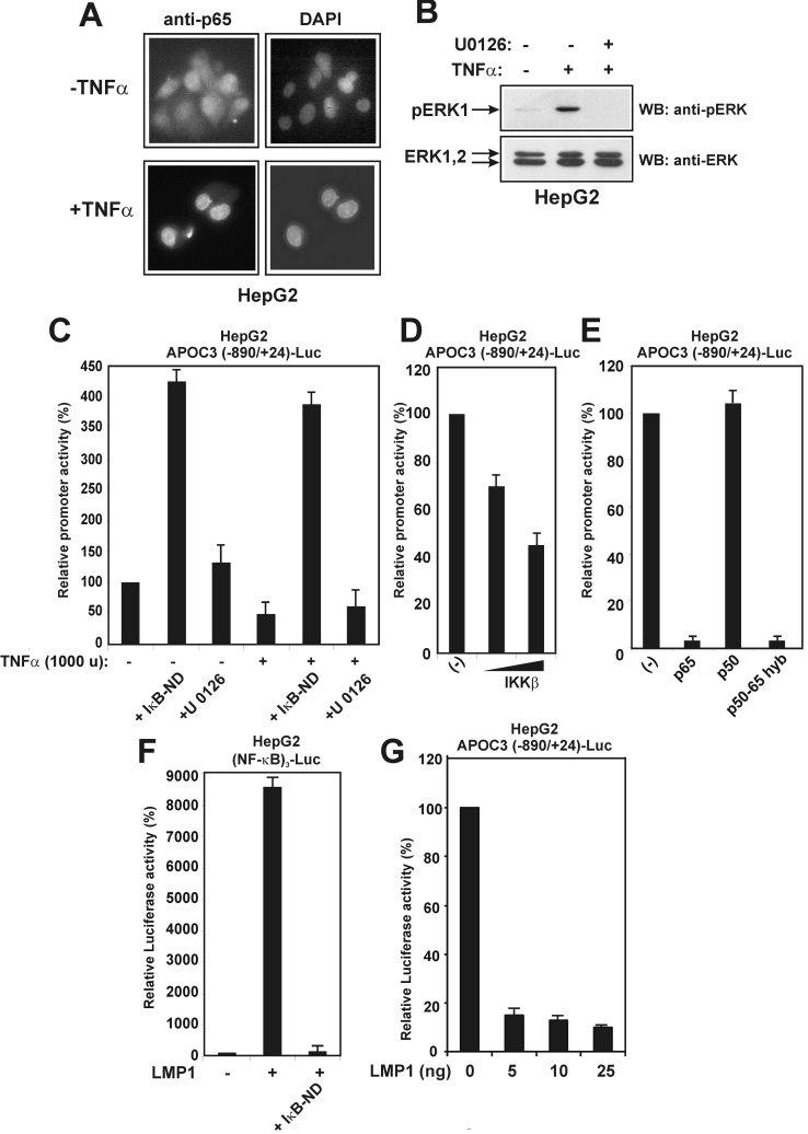
Figure 2. Inhibition of APOC3 promoter activity by TNFα is mediated by NF-κB.
(A) TNFα activates NF-κB in HepG2 cells. HepG2 cells were serum-starved for 16 h and treated with TNFα (1000 units) for 4 h or left untreated. The intracellular distribution of the p65 subunit of NF-κB was examined by indirect immunofluorescence using an anti-p65 antibody followed by a secondary FITC-conjugated antibody. Nuclei were stained with DAPI (4′,6-diamidino-2-phenylindole). (B) TNFα activates the MEK1/ERK pathway in HepG2 cells. HepG2 cells were pretreated with the MEK1 inhibitor U0126 (10 μM) for 24 h, followed by a short treatment with TNFα (1000 units) for 15 min. Cell extracts were analysed by Western blotting for pERK or total ERK using the corresponding anti-ERK antibodies followed by secondary horseradish-peroxidase-conjugated antibodies. Arrows show the position of pERK and ERK1/2 proteins. (C) HepG2 cells were transiently transfected with the APOC3 (−890/+24)-Luc reporter plasmid (2 μg) and treated with TNFα (1000 units) in the presence or in the absence of an expression vector for the non-degradable form of IκBα, IκB-ND (2.0 μg), or the MEK1 inhibitor U0126 (10 μM) for 24 h. Cell extracts were analysed for luciferase activity. Normalized relative APOC3 promoter activity is shown as a histogram. (D) HepG2 cells were transiently transfected with the APOC3 (−890/+24)-Luc reporter plasmid (2 μg), along with increasing concentrations of an expression vector for IKKβ (0, 1.0 and 2.0 μg). Cell extracts were analysed for luciferase activity. Normalized relative APOC3 promoter activity is shown as a histogram. (E) HepG2 cells were transiently transfected with the APOC3 (−890/+24)-Luc reporter plasmid (2 μg), along with expression vectors for p65, p50 or a p50/p65 hyb protein (2.0 μg each). Cell extracts were analysed for luciferase activity. Normalized relative APOC3 promoter activity is shown as a histogram. (F) HepG2 cells were transiently transfected with the (NF-κB)3-Luc reporter plasmid (2 μg), along with an expression vector for LMP1 (25 ng) in the presence or in the absence of an expression vector for IκB-ND (2.0 μg). Cell extracts were analysed for luciferase activity. Normalized relative APOC3 promoter activity is shown as a histogram. (G) HepG2 cells were transiently transfected with the APOC3 (−890/+24)-Luc reporter plasmid (2 μg), along with increasing concentrations of an expression vector for LMP1 (0, 5, 10 and 25 ng). Cell extracts were analysed for luciferase activity. Normalized relative APOC3 promoter activity is shown as a histogram.

