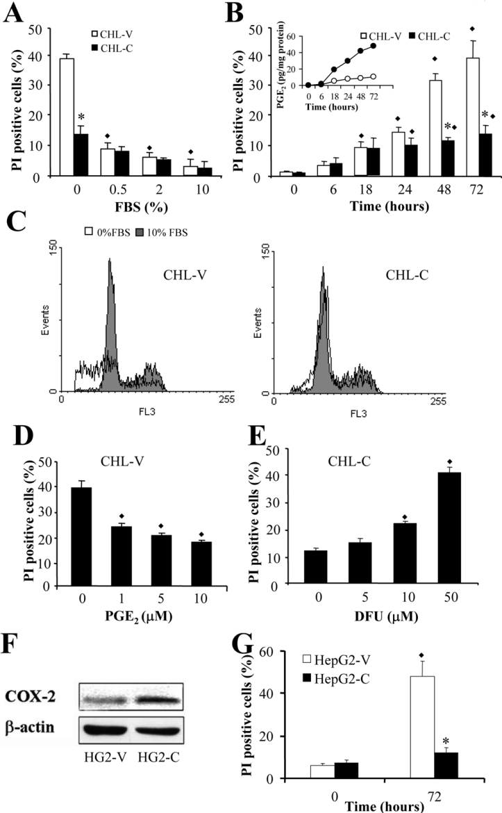
Figure 3. COX-2 expression inhibits PI staining in liver cells.
Cells resuspended in PBS were PI-stained, and cell-cycle analysis was determined by flow cytometry. (A) Effect of FBS concentration on the percentage of apoptotic CHL-V and CHL-C cells after 72 h of culture. (B) Time-dependent apoptosis and PGE2 concentrations in CHL-V and CHL-C cells cultured without FBS. The inset shows the level of PGE2 in each cell type over the same time period. (C) Representative DNA histograms and G1 profiles showing the PI staining from CHL-V and CHL-C cells cultured at 0 and 10% (v/v) FBS for 72 h. (D) Effect of exogenous PGE2 on the percentage of apoptotic cells. (E) Effect of DFU on the percentage of apoptotic CHL-C cells (no FBS, 72 h of culture). (F) Western blot analysis of COX-2 expression in HepG2-V and HepG2-C. (G) Apoptosis of HepG2-V and HepG2-C cells without FBS after 72 h of culture. Results are means±S.D. for four independent experiments. *P<0.05 compared with paired CHL-V cells. ◆P<0.05 compared with cells at zero time (B) or untreated cells (D and E).

