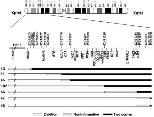
Figure 2. .
Detailed schematic representation of the 3′ deletion limits of probands 10P and 43–48, which extend beyond the PAR1 into X-specific regions. These samples correspond to one male (proband 43), five sporadic females (44, 45, 10P, 47, and 48), and proband 46, a familial case from a family in which only affected females were observed. Blackened areas indicate the presence of two copies of the marker or SNP, unblackened areas indicate the presence of a deletion, and shaded areas indicate the noninformative areas where the breakpoints are located. Localization of the microsatellite markers and the genes located within the deletions are indicated by vertical lines above and below the line, respectively.

