Figure 3. .
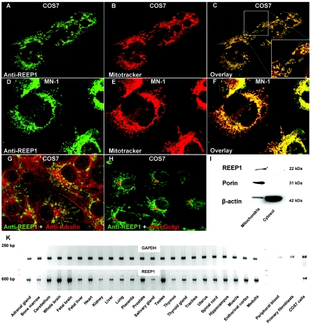
REEP1 localized to mitochondria. A–F, Immunohistochemistry with two different REEP1 antibodies revealed colocalization with the marker Mitotracker Red. COS7 (kidney) and MN-1 (motor neuron) cells were maintained in Dulbecco’s modified Eagle medium with 10% fetal bovine serum and were incubated at 37°C supplemented with 5% CO2. Cells were observed by confocal laser scan microscopy (Visitech model VT-Infinity). G and H, No colocalization was observed with microtubules and Golgi. I, COS7 cells were homogenized and mitochondria were separated from the cytosolic fraction by centrifugation. The REEP1 antibodies were probed against COS7 cell lysate on a western blot and showed a band of the expected size in the mitochondrial fraction. The blot was reprobed with porin, which constitutes a mitochondrial membrane protein, and β-actin. Note that the cytosolic fraction was highly enriched, as shown by the β-actin bands. J, RT-PCR on a series of tissues revealed ubiquitous expression. REEP1 was not expressed in peripheral blood and fibroblasts derived from a skin biopsy.

