
Figure 2.
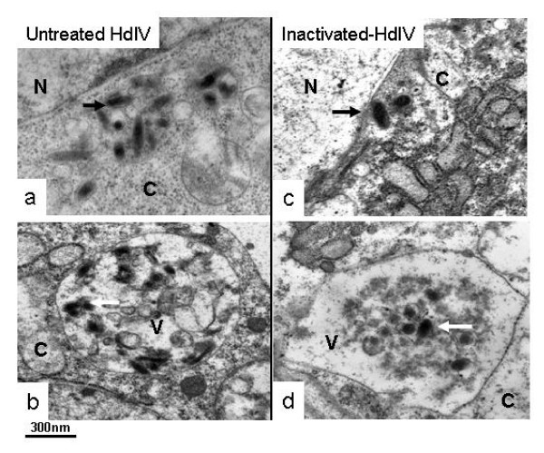
Infection of Spodoptera frugiperda hemocytes after infection of untreated HdIV (left panels, a and b) and heat-inactivated HdIV (right panels, c and d) by transmission electronic observations. For both control and heat treatment, HdIV particles can be observed within the cytoplasm of hemocytes (black arrows, a and c) and in vacuoles (white arrows, b and d). C: cytoplasm; N: nucleus; V: vacuole.