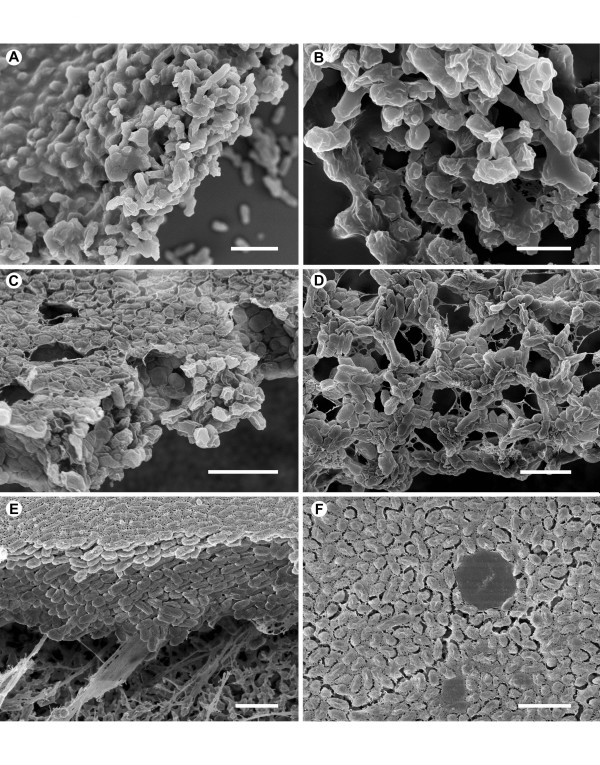
Figure 1.

Nontypeable Haemophilus influenzae biofilm imaged via scanning electron microscopy. Scanning electron micrographs of NTHi biofilms formed under different growth conditions. A and B) Sterile glass coverslips were covered with a suspension of NTHi in BHI broth. After 24 hr, the coverslips were prepared for SEM examination. (A) Large flat mats of bacteria embedded in an amorphous extracellular matrix were found attached to the glass surface. Scale bar = 2 μm. (B) The individual NTHi are covered in an amorphous layer that conceals the bacterial surface. Scale bar = 1 μm. C and D) Suspensions of NTHi in BHI broth were placed onto sterile Anopore insert filters that were mounted on chocolate agar. Once the NTHi biofilms had formed, after 24 hr incubation, on the upper surface of the filters at the air/liquid interface, the inserts were placed in culture dishes containing sufficient sterile culture medium to exert a positive upward pressure on the bottom of the biofilm, and left for a subsequent 24 hr. (C) The surface of the insert filter is covered with a flat mat consisting of NTHi closely attached to each other. Channels and pockets freee of bacteria have formed within the mat of bacteria. Scale bar = 2 μm. (D) In some orientations, it is possible to see the channels running between the aggregates of bacteria and through the mat. Scale bar = 2 μm. E and F) NTHi biofilms grown on Millipore filters. Sterile Millipore filters were placed onto chocolate agar plates and inoculated with sufficient NTHi in BHI broth to cover the surface at a density of 0.3 bacteria per 10 μmP2P. The filters were incubated for 24 hr with the upper surface exposed to air, and prepared for SEM examination. (E) The NTHi formed thick biofilms with the base firmly attached to the filter substrate. Scale bar = 2 μm. (F) The top surface of the NTHi biofilm, that had been exposed to air, was covered with a thin film of extracellular matrix. In some instances, the matrix formed a film over regions that resembled bacteria-free pockets. Scale bar = 2 μm.
