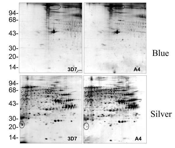
Figure 1.
2D electrophoresis profile of Coomassie Blue (upper) and silver (lower) stained iRBC ghosts from 3D7 (left) and A4 (right). The first dimension was run on pH4-7 IEF strips followed by 12.5% SDS-PAGE. The two major changes in the protein profiles are ringed (see text for details). High quality tif files showing the original 2D gels are available for 3D7 iRBC ghosts stained with Coomassie Blue (Additional file 1) and Silver (Additional file 2), and for A4 iRBC ghosts stained with Coomassie Blue (Additional file 3) and silver (Additional file 4).

