
Figure 5.
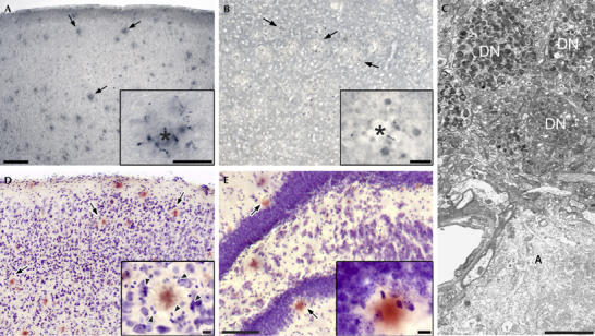
Amyloid-associated pathology in 8-month-old APPPS1-21 mice. (A) Immunostaining (AT8 antibody) shows hyperphosphorylated tau-positive neurites surrounding amyloid deposits (arrows) in the neocortex. The inset shows a plaque (asterisk) and abnormal neurites in higher magnification. (B) Synaptophysin-positive dystrophic terminals and boutons around amyloid plaques in layer III of the frontal cortex. (C) Electron micrograph of dystrophic neurites (DN) surrounding an amyloid plaque (A) in neocortical layer II. (D,E) Cresyl violet and congo red staining in the neocortex (D) reveals a ring of glial cells (arrowheads in the inset) surrounding the amyloid plaque. Neurons appear physically displaced but look normal in the periphery of the plaques. In the dentate gyrus (E), a thinning of the granule cell layer in the vicinity of amyloid plaques is apparent (arrows) although neurons with an unequivocally dying phenotype are rarely observed. Scale bars: 100 μm (A,B,D); 50 μm (E); 20 μm (insets); 5 μm (C).