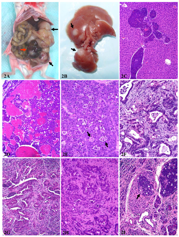
Figure 2.
Alterations of the pancreas from Ela-myc transgenic mice. A: a photo showing a huge nodular pancreatic tumor (arrows). Note that one tumor nodule is in red color while other tumor nodules are in white color. B: liver metastases (arrows) of a pancreatic tumor. C: histological examination confirming that the liver tumors are pancreatic origin (acinar cell carcinoma). D: a typical histology of acinar cell carcinoma that shows red color macroscopically. E: a typical histology of the acinar cell carcinoma that shows white color macroscopically. Note that there are many apoptotic cells that are organized in clusters, coined as "death cell islands" (arrows). F: a typical area of mixed acinar and ductal adenocarcinomas. G: a pancreatic ductal adenocarcinoma. Note that the tumor contains abundant stroma. H: another typical ductal adenocarcinoma. I: an acinar cell carcinoma within an islet (arrow).

