Figure 2.
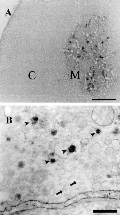
Adult rat adrenal gland reacted with antireelin antibody G10. (A) Low-power photo. C, adrenal cortex; M, adrenal medulla. Scattered clusters of chromaffin cells are positive, whereas the cortex is negative. (Bar = 0.4 mm.) (B) Immunoelectron microscopy of chromaffin cells. Dense reelin-like immunoreactivity is observed in a subset of chromaffin granules. Arrowheads show reelin-positive granules; arrows show reelin-negative granules. Asterisks indicate reelin-like immunoreactivity within the extracellular space. (Bar = 0.5 μm.)

