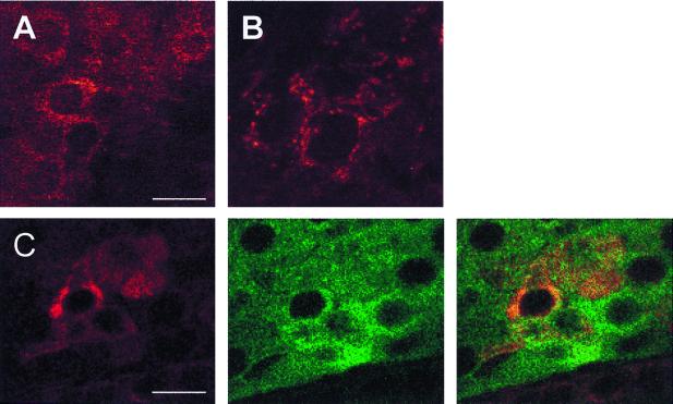
Figure 3.
Reelin-like immunostaining in adult rat adrenal medulla and pituitary pars intermedia viewed by confocal microscopy. (A) Adrenal chromaffin cells, stained with antireelin antibody 142. (Bar = 20 μm.) (B) Pituitary pars intermedia, stained with antireelin antibody G10. (C) Double labeling of pituitary pars intermedia cells for reelin and α-MSH. (Left) Antibody G10. (Bar = 10 μm.) (Center) Anti-MSH. (Right) Overlay shows that a subset of the α-MSH-positive cells express reelin.

