Figure 6.
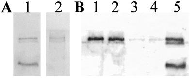
Reelin-immunoreactive bands in liver in situ and in vitro. (A) Adult rat liver extracted and Western-blotted by using antireelin antibody G10. Lane 1: calibration standard (cerebellar granule cell-conditioned medium). Lane 2: liver extract. (B) Adult rat liver was dissociated into single cells by perfusion with collagenase, followed by sieving to remove tissue clumps. Cells were plated in collagen-coated dishes at ≈80% confluence, allowed to recover in full feeding medium containing serum for 4 hr, rinsed five times in minimal serum-free medium (Williams' E medium with BSA, glutamine, and antibiotics), and then incubated with this medium with changes immediately, after 5 hr, and overnight. Conditioned medium was harvested, spun to remove cellular debris, and assayed for reelin by Western blotting. Lanes: 1 and 2, conditioned medium collected overnight from duplicate culture wells shows that reelin was released primarily as the full-length, 420-kDa reelin gene product; 3 and 4, same as lanes 1 and 2 but incubated in the presence of dexamethasone (2 μM); 5, calibration standard (cerebellar granule cell-conditioned medium). As a control, no reelin-like immunoreactivity was observed in the conditioned medium incubated with liver cells momentarily, and more reelin was observed after overnight incubation than after 5 hr, indicating that the release of reelin was time-dependent (not shown). Extracts of the liver cells examined after overnight incubation also contained a predominant, 420-kDa reelin band that was decreased in the presence of dexamethasone (not shown).

