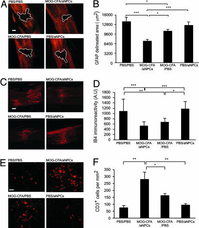
Fig. 3.
Local changes in immune activity correlate with tissue preservation. On the day of SCI, mice were vaccinated with MOG peptide emulsified in CFA containing 0.5 mg/ml M. tuberculosis. Seven days later, the vaccinated and control mice were either transplanted with aNPCs or injected with PBS. Their spinal cords were excised 1 week after cell transplantation. (A) Representative micrographs showing GFAP staining of spinal cords from mice treated with MOG-CFA/aNPC, MOG-CFA/PBS, PBS/aNPC, or PBS/PBS are shown. (B) Quantification of the area delineated by GFAP staining. (C) Representative micrographs of IB4-stained areas. (D) Quantification of IB4 immunoreactivity. (E) Representative micrographs of CD3 staining, identifying infiltrating T cells in the area surrounding the site of injury. (F) Quantification of CD3+ cells in the area surrounding the site of injury (n = 4 for MOG-CFA/aNPC and n = 3 for MOG-CFA/PBS, PBS/aNPC, and PBS/PBS; ∗, P < 0.05; ∗∗, P < 0.01; ∗∗∗, P < 0.001, ANOVA). Data are means ± SEM. (Scale bar, 100 μm in A and C; 20 μm in E.)

