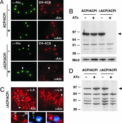
Fig. 5.
Loss of apicoplast FAS II abolishes lipoylation of apicoplast but not mitochondrial enzymes. (A and B) Immunofluorescence (A) and Western blot (B) analysis of parent and mutant cells with antibody to lipoylated PDH-E2 (2H-4C8). ATc treatment of the mutant abolishes PDH labeling (arrow). An antibody to the T. gondii apicoplast histone-like protein (Hu protein; S. Vaishnava and B.S., unpublished observations) served as apicoplast marker, and an antibody to MIC2 served as loading control. (C and D) Immunofluorescence (C) and Western blot (D) analysis using a polyclonal serum reactive to all lipoylated enzymes localized to the plastid (P) and the mitochondrion (M). Note loss of PDH labeling (black arrowhead) and persistence of mitochondrial labeling (white arrowhead).

