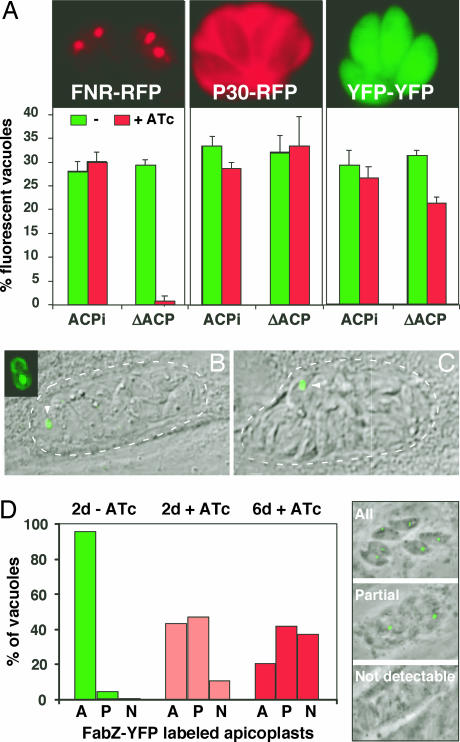
Fig. 6.
Depletion of ACP leads to defects in apicoplast morphology and biogenesis. (A) ACP/ACPi and ΔACP/ACPi cells were grown for 6 days in the presence or absence of ATc and then transiently transfected with plasmids, resulting in the expression of FNR-RFP (apicoplast), P30-RFP (dense granules and parasitophorous vacuole), and YFP-YFP (cytoplasm) before infection of coverslip cultures. After 24 h parasites were scored for fluorescent protein expression. (B and C) A ΔACP/ACPi line stably expressing the apicoplast marker FabZ-YFP was treated with ATc for 3 days and imaged in vivo. Vacuoles with a single or a few large apicoplasts were frequently observed (arrowhead; parasitophorous vacuoles are indicated by dotted lines). (D) The same line was treated with ATc for 0, 2, and 6 days, and plastid morphology was scored by using the three categories indicated (A, all parasites in a vacuole show a fluorescent apicoplast; P, one or few apicoplasts per vacuole; N, no apicoplast labeling detected).

