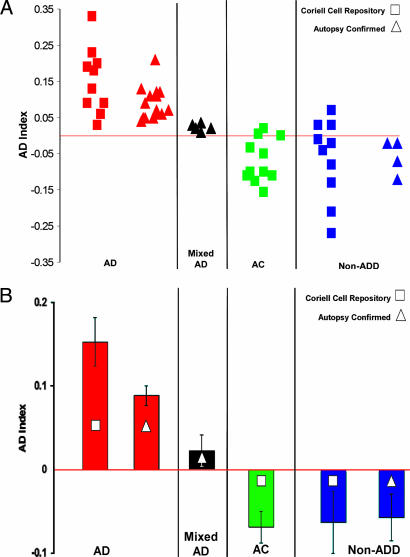
Fig. 3.
AD index values for Erk1/2 phosphorylation in human fibroblasts. (A) The AD index was plotted for cells of patients from four different categories: (i) AD, (ii) mixed AD/PD/DLV (mixed diagnosis of AD, Parkinson’s disease, and Lewy body disease) as confirmed by autopsy, (iii) age-matched control (AC), and (iv) non-AD dementia (non-ADD; i.e., Parkinson’s disease and Huntington’s disease) for Coriell Cell Repository and autopsy-confirmed cell lines. For all AD diagnoses, the AD index had positive values. (B) The mean indices for AD cases were positive and higher than those for ACs and non-ADD patients. Mixed (AD/PD/DLV) autopsy patients had lower but still positive AD index values. Significance levels between groups were as follows: P < 0.00001 for AD vs. AC cells from the Coriell Cell Repository, P < 0.0001 for AD vs. non-ADD cells from autopsy-confirmed cases, and P < 0.002 for AD vs. mixed AD diagnosis cases.

