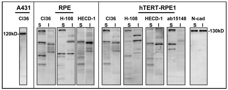
Figure 5.

E-cadherin Western blots of protein extracts from RPE cultures at 30 hours after plating showing banding patterns for cells from a human donor, aged 71 years in passage 5 (RPE) and for the hTERT-RPE1 cell line. Different E-cadherin antibodies were used (cl36, H-108, HECD-1, and ab15148) to blot Triton detergent-soluble (S) and -insoluble (I) fractions. Also shown are the migration positions of E-cadherin (120 kDa) in a whole-cell extract of A431 cells and of N-cadherin (130 kDa) in soluble and insoluble fractions of hTERT-RPE1 cells.
