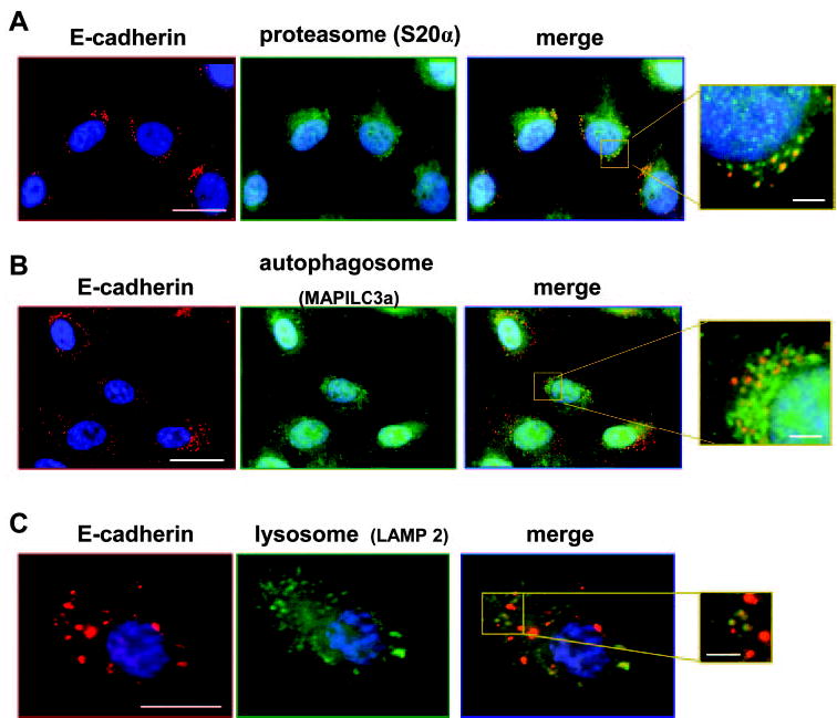
Figure 8.

hTERT-RPE-1 cells at 30 hours after plating costained for E-cadherin (clone 36, red) and organelle markers (green) for (A) the proteasome (S20α), (B) the autophagosome (MAPILC3a), and (C) the lysosome (LAMP2). As shown in the enlarged merged images, a subset of the E-cadherin-stained granules colocalized with each organelle (yellow). Scale bars: 20 μm (low magnification images), 2 μm (enlargements).
