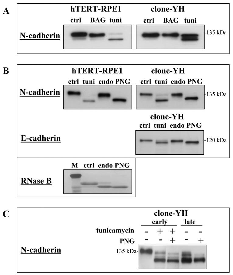
Figure 5.

Cadherin glycosylation in hTERT-RPE1 and clone-YH cells. (A) Immunoblot analysis of N-cadherin after treatment of cells in early culture with inhibitors of N-glycosylation (0.5 μg/mL tunicamycin [tuni] for 18 hours) or O-glycosylation (2 mM benzyl-N-acetyl-α-galactosaminide [BAG] for 24 hours). Only tunicamycin produces a reduction in cadherin mass, although the extent of deglycosylation varied among experiments. (B) Cadherin immunoblots of early cultures that were untreated (control [ctrl]) or treated with tunicamycin (tuni). Extracts of control cultures were subsequently incubated with endoglycosidase H (endo) or peptide N-glycosidase F (PNG). Only PNG shows an effect. Gly-cosidase treatment of RNase B is shown as a positive control for the digestion protocols; M indicates molecular mass standard. (C) N-cadherin immunoblot of early and late clone-YH cultures. Early cultures were untreated or treated with tunicamycin. Extracts of early and late cultures were incubated without or with PNG. N-cadherin in tunicamycin treated early cultures comigrates with N-cadherin in late cultures. The migration position of N-cadherin in late cultures is unchanged by PNG treatment.
