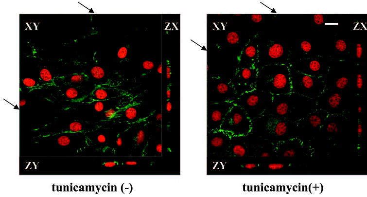
Figure 8.

Confocal microscopy of clone-YH without or with tunicamycin treatment, showing the epithelioid phenotype and zonular distribution of N-cadherin after tunicamycin. The arrows indicate the faint lines on the composite en face images, which show the position of the cross-sectional ZX and ZY scans. Nuclei are counterstained with SYTOX Orange. Scale bar, 10 μm.
