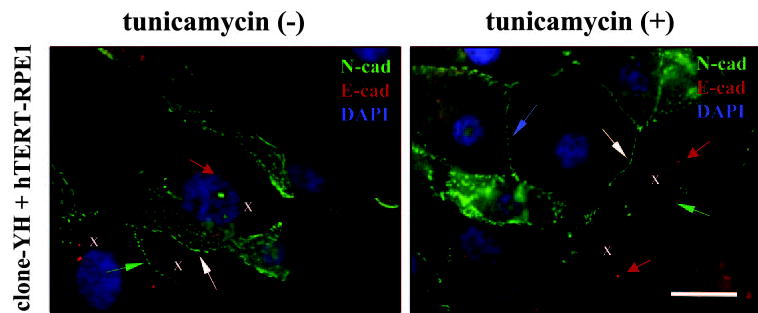
Figure 9.

Early cocultures of clone-YH and hTERT-RPE1 without or with tunicamycin treatment co-stained for N-cadherin (N-cad, green), E-cadherin (E-cad, red), and DAPI (for nuclei, blue). hTERT-RPE1 cells were identified by their centrosomal-associated E-cadherin immunoreactivity (red arrows); some hTERT-RPE1 cells in the images are indicated by Xs. N-cadherin can be seen localizing to homotypic junctions between clone-YH cells (blue arrow) or between hTERT-RPE1 cells (green arrows). The junctions between hTERT-RPE1 in the treated cultures are poorly developed. N-cadherin also localizes prominently to heterotypic junctions between the two cell types (white arrows). Scale bar, 20 μm.
