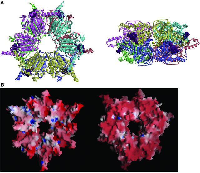
Fig. 2. 12S hexamer. (A) Ribbon diagram viewed down (left) and perpendicular to (right) the 3-fold rotation axis. Monomers are in different colors, MMCoA molecules are in purple space-filling representation, and cadmium ions are yellow spheres. (B) Electrostatic surface of the trimer stacking face (left) and solvent-exposed surface (right).

An official website of the United States government
Here's how you know
Official websites use .gov
A
.gov website belongs to an official
government organization in the United States.
Secure .gov websites use HTTPS
A lock (
) or https:// means you've safely
connected to the .gov website. Share sensitive
information only on official, secure websites.
